Migraines & chronic headaches / Auckland & Christchurch
Migraines And
Chronic Headaches
For sufferers who have tried everything.
Could the Source of Your Migraines Be Trapped Deep Inside Your Head?
If medications and therapies haven’t brought lasting relief, it’s often because they only treat the blood vessels, not the mechanical cause. Deep inside your skull, the central bones form a bridge where key nerves pass through. When those bones shift or lock up, they pull on sensitive membranes and irritate the main nerve pathway that triggers severe migraine throbbing.
Nasal RX works directly through your natural nasal passages to release that structural tension, calming the nerve signals and treating the root cause of your pain.
Book a Structural Assessment Explore the Trigeminal PathwayWhat is Nasal RX for chronic migraines and headaches?
Nasal RX is a non-surgical endonasal procedure offered in Auckland and Christchurch that addresses structural tension in the cranial base associated with chronic headaches and migraines. Suitability is decided at an individual assessment, and results vary from person to person.
Clinical overview / The trigemino-vascular cascade
The Trigemino-Vascular Cascade
Migraines are not merely blood vessel dilations. They are frequently triggered by physical mechanical traction along dural membranes where the trigeminal nerve passes through the central sphenoid bone. When cranial sutures lock, dural strain sensitises nerve pathways, prompting neuropeptides such as CGRP to drive vascular pain signals.
Trigeminal pathway simulator
Trigeminal Ganglion Pressure Simulator
Toggle dural tension states to visualise nerve compression at the sphenoid base.
High mechanical strain sensitising dural nerves and driving neurovascular throbbing.
Introduction / The persistent cycle
Exhausting Cycle
If you live with chronic migraines, you know the exhausting cycle all too well. You have likely tried over-the-counter painkillers, prescription triptans, monthly CGRP injections, preventive medications, diets, dark rooms, and endless lifestyle adjustments. You may have even had brain scans (like CTs or MRIs) that came back “completely normal,” leaving you wondering why your head still feels trapped in a vice.
When Standard Treatments Fail
When standard treatments fail to provide lasting relief, it is often not because your pain is “all in your head.” It is because standard treatments focus almost entirely on brain chemistry and blood vessels, while overlooking a major physical driver of chronic head and neck pain: mechanical tension across the outer lining of your brain (the Dura Mater) and its skull anchor points.
This page explains why chemical treatments alone often leave physical pain intact, how locked skull seams (sutures) create constant dural tension, and how the Nasal RX procedure uses gentle internal micro-mobilisation to release this deep physical strain at its source.
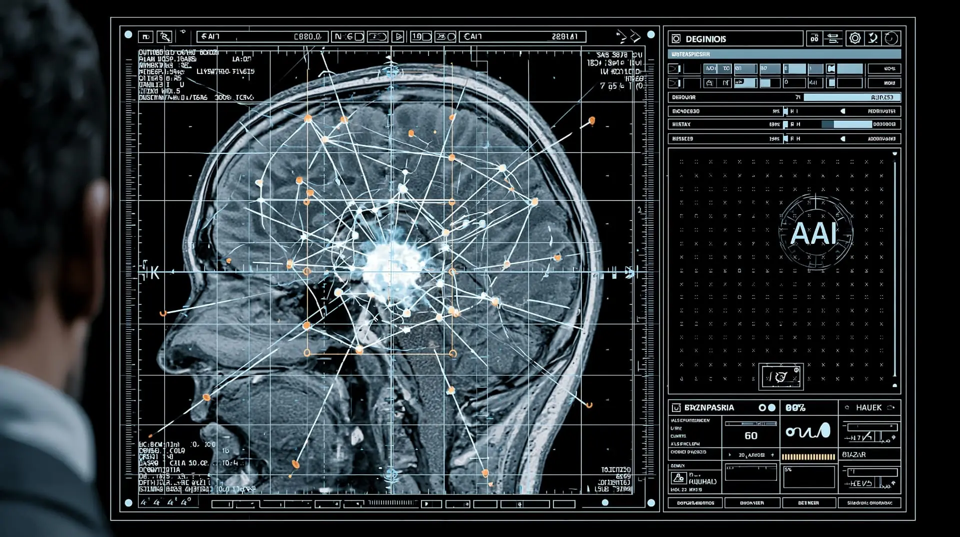 Plate 01 / Cranial scan
01 / Clinical context
The Headache &
Neck Pain Link
Clinical proof
A major medical meta-analysis published in Cephalalgia (Danish Headache Center / Harvard Medical School) reviewed 24 clinical studies and found:
- 77.0%
- of Episodic Migraine sufferers have co-occurring mechanical neck strain.
- 87.0%
- of Chronic Migraine sufferers have co-occurring mechanical neck strain.
- 12×
- Neck pain is more common in Migraine patients than non-Headache controls.
Nasal RX Neck Protocol
Recognising the direct neurological connection between the skull base and the upper spine, Nasal RX protocols encompass full cervical structural screening. We thoroughly evaluate upper neck alignment and resolve biomechanical imbalances, ensuring both the central cranial base and contributing cervical factors are comprehensively addressed.
Comparing Treatment Models
A side-by-side comparison of the standard pharmaceutical / vascular model and the Nasal RX structural protocol.
Treatment Modality
- Pharmaceutical / Vascular Model
- Daily pills, monthly self-injections, or dark-room resting.
- Nasal RX Structural Protocol
- Non-surgical, gentle endonasal inflation micro-mobilisation.
Primary Mechanism
- Pharmaceutical / Vascular Model
- Chemical receptor blockade or blood vessel narrowing (triptans / CGRP).
- Nasal RX Structural Protocol
- Physical decompression of skull base dural attachments.
Key Anatomical Target
- Pharmaceutical / Vascular Model
- Chemical receptors (CGRP, serotonin) in blood vessels.
- Nasal RX Structural Protocol
- Sphenoid bone, temporal ridges, and internal dural partitions.
Neck Pain Connection
- Pharmaceutical / Vascular Model
- Viewed as an unrelated side effect or muscle stress.
- Nasal RX Structural Protocol
- Recognised as a direct trigemino-cervical reflex muscle spasm.
Eye & Light Sensitivity
- Pharmaceutical / Vascular Model
- Treated with dark rooms, filters, or systemic drugs.
- Nasal RX Structural Protocol
- Addressed by releasing dural-scleral traction behind the eye.
Long-Term Impact
- Pharmaceutical / Vascular Model
- Requires repeated dosing; rebound headaches possible.
- Nasal RX Structural Protocol
- Restores baseline structural and dural equilibrium.
| Clinical Feature | Pharmaceutical / Vascular Model | Nasal RX Structural Protocol |
|---|---|---|
| Treatment Modality | Daily pills, monthly self-injections, or dark-room resting. | Non-surgical, gentle endonasal inflation micro-mobilisation. |
| Primary Mechanism | Chemical receptor blockade or blood vessel narrowing (triptans / CGRP). | Physical decompression of skull base dural attachments. |
| Key Anatomical Target | Chemical receptors (CGRP, serotonin) in blood vessels. | Sphenoid bone, temporal ridges, and internal dural partitions. |
| Neck Pain Connection | Viewed as an unrelated side effect or muscle stress. | Recognised as a direct trigemino-cervical reflex muscle spasm. |
| Eye & Light Sensitivity | Treated with dark rooms, filters, or systemic drugs. | Addressed by releasing dural-scleral traction behind the eye. |
| Long-Term Impact | Requires repeated dosing; rebound headaches possible. | Restores baseline structural and dural equilibrium. |
Clinical deep-dive library
The Full Deep-Dive Library
All long-form clinical content is preserved below. Tap any heading to expand the full anatomy, mechanics, evidence and references.
Did you know?
Your Brain Feels No Pain. Your Brain’s Lining Does
Millions of chronic headache sufferers are surprised to learn that brain tissue has zero pain receptors. The deep, vice-like pressure you feel stems directly from mechanical tension across the Dura Mater - the structural lining anchored to your central skull base.
Acting like internal tension wires, nerves embedded in this dural sheet fire pain signals the moment mechanical strain stretches or twists its skull base anchors.
Why Nasal RX Is the Preferred Non-Surgical Strategy
Because the central sphenoid bone forms the main structural anchor for your internal dural lining, addressing this pain requires an internal structural approach. The Nasal RX procedure works from within the nasal vault to gently micro-mobilise restricted sphenoid seams. This releases stored dural tension, calms overactive head nerves, and interrupts the mechanical pain cycle without surgery or ongoing pharmaceutical dependence.
Chemical Band-Aids vs. Physical Pulling Forces
Most modern headache drugs-such as over-the-counter NSAIDs, prescription triptans, and CGRP receptor blockers-are designed to change chemical signaling. They work by blocking pain chemicals or narrowing swollen blood vessels.
While chemical blocking agents can provide helpful temporary relief during an acute attack, they do not alter physical mechanical pulling across your skull base. If your skull bones are slightly jammed or twisted, the physical stretch on your Dura Mater continues to pull like a tight rubber band. As soon as the medication wears off, the physical stretch reactivates your pain nerves.
Article 1: Why Migraine Medications & Scans Leave Physical Strain Unaddressed.
Article 11: Medication-Overuse Rebound Headaches vs. Physical Dural Rebound.
How Strong Is Your Brain’s Lining?
Biomechanical testing using specialised tensile testing equipment proves that the human Dura Mater is an exceptionally tough, elastic sheet. It has an elastic stiffness (Young’s Modulus) measuring between 28 MPa and 86 MPa, with high tensile strength.
The collagen fibers inside the dural lining are arranged in crisscross layers. This structure makes the Dura Mater anisotropic, meaning its strength and stretchiness depend heavily on the direction in which it is pulled:
- Directional Stiffness
- The dural lining is significantly stiffer when pulled lengthwise (sagittal) than when pulled sideways (transverse).
- Sensitivity to Structural Twisting
- Because the lining is stiff and direction-dependent, even subtle misalignments in your skull bones exert uneven pulling forces across internal brain walls.
- Continuous Nerve Firing
- Physical stretch receptors on dural nerve endings open physically under tension, firing electrical pain signals as long as the mechanical stretch remains.
The Internal Tug-of-War
How Locked Skull Anchors Pull Your Brain's Protective Lining Taut
High tension
Restricted skull seams pull internal brain linings taut like an over-stretched drum skin, firing continuous pain signals.
- Front Anchor
- Behind EyesEases deep orbital pressure.
- Side Anchor
- Temples & JawEases temple throbbing & jaw strain.
- Back Anchor
- Skull BaseRelieves upper neck knots & stiffness.
Internal Brain Partitions & Skull Attachment Points
The meningeal layer of the Dura Mater folds inward to create internal support walls that keep your brain stable during movement. The falx cerebri anchors at the crista galli behind the nose and blends into the dural roof at the back of the skull. The tentorium cerebelli connects to the sphenoid, temporal and occipital bones.
Your brain is held stable by internal dural partitions that anchor directly onto your skull bones:
The Vertical Wall (Falx Cerebri): Anchors right behind your nose at the crista galli and extends to the back of the skull.
The Horizontal Floor (Tentorium Cerebelli): Straps directly across your sphenoid, temporal, and occipital bones.
When mechanical restrictions jam these skull base bones, these internal walls twist and pull taut. Nasal RX works directly from within the nasal passageway to gently mobilise the sphenoid anchor, easing this internal structural pull.
Skull base dural anchor map
Crista Galli & Sphenoid Lesser Wings
Sphenoid Clinoid Processes & Sella Turcica
Petrous Ridge of Temporal Bone
Transverse Sulci of Occipital Bone
Anatomical map showing how internal dural walls anchor directly onto the sphenoid and temporal bones, forming a continuous internal tension system.
When cranial seams become restricted or jammed-whether from past head bumps, dental work, jaw clenching, or poor posture-these internal dural walls are pulled taut like a stretched trampoline.
Article 3: How Locked Skull Bones & Sutures Create Deep Vise-Like Head Pressure.
Article 6: One-Sided Eye Pain & Asymmetric Dural Traction.
Why Headaches and Neck Pain Show Up Together
If you experience neck tightness during a migraine, you are not alone. Clinical research shows that up to 87% of chronic migraineurs suffer from severe upper neck pain.
This happens because sensory nerves from your skull lining (Trigeminal Nerve - CN V1) and sensory nerves from your top three neck joints (C1, C2, and C3) feed into the exact same neurological “junction box” in your upper spinal cord: the trigeminocervical nucleus.
Because these nerves share a single processing hub, your brain often cannot distinguish between physical strain in your skull lining and joint tightness in your neck. Physical dural tension at your skull base easily feels like a throbbing pain behind your eye, while upper neck joint tightness can set off a full-blown migraine.
Automatic Muscle Spasms in Your Neck
- Protective Head Guarding
- Irritated head nerves automatically activate motor nerve cells controlling your neck muscles, particularly the sternocleidomastoid (SCM) and suboccipital muscles.
- Reflex SCM Tightness
- The SCM muscle attaches to the mastoid bone right behind your ear. Dural stretch signals can command the SCM muscle to contract in defense.
- Sustained Muscle Guarding
- This automatic reflex loop keeps your neck muscles locked in painful knots, explaining why neck stiffness is so difficult to rub or stretch away.
The reflex muscle-guarding cycle
- 1
Physical Stretch on Skull Base Anchors (Sphenoid/Temporal)
- 2
Head Sensory Nerves Fire Pain Signals (CN V1)
- 3
Signals Enter the Shared Nerve Junction Box
- 4
Automatic Reflex Triggers Neck Motor Nerves
- 5
Neck Muscles (SCM) Contract to Protect the Head
The trigemino-cervical reflex loop showing how head lining strain triggers automatic muscle guarding in the neck muscles.
Ref. 2: Why Upper Neck Stiffness & Base-of-Skull Knots Always Accompany Migraines.
Ref. 9: Morning Migraines & Waking Up in Pain.
Why You Feel Crushing Pain Behind Your Eyes & Why Weather Drops Set Off Attacks
If your headaches cause deep, aching pain behind your eyeballs, extreme light sensitivity (photophobia), or severe flares during weather shifts, your symptoms are not random. They stem from a direct physical connection between your eyes, your immune system, and the internal protective lining of your brain (the Dura Mater).
The Direct Eye-to-Dura Pathway
- 01
Central Sphenoid Bone Anchor
Behind the nose and eye sockets.
- 02
Physical Stretch on Dura Mater
Skull base anchors pull the dural lining taut.
- 03
Transmits Along the Optic Nerve Sheath
Strain travels the nerve sheath toward the eye.
- 04
Fuses Into the Back of the Eyeball
The sheath anchors directly into the sclera.
Part 1: How Physical Stretch Reaches Your Eyes
The outer sheath surrounding your optic nerve is a direct extension of your brain’s dural lining, fusing tightly into the back wall (sclera) of your eyeball.
Because the main dural roof anchors directly onto the central sphenoid bone right behind your nose and eye sockets:
- Tension Transmission
- Any mechanical strain or twisting across central sphenoid anchors pulls directly along the optic nerve sheath, creating an actual physical tug at the back of your eye.
- Photophobia (Light Sensitivity)
- Pain nerves (CN V1) embedded in this eye-dural sheath become pre-sensitised by physical stretch. When light enters your eye, these pre-irritated nerves cause normal room light to feel painful.
Part 2: The Mast Cell Inflammation Cycle
The Dura Mater is packed with specialised immune cells called dural mast cells, sitting right beside your pain nerves.
When skull base anchors pull the dural sheet taut, this physical stretch causes mast cells to degranulate, releasing inflammatory chemicals like histamine and CGRP. This local inflammation lowers your nerves’ activation threshold, making your head highly vulnerable to everyday triggers:
- Barometric Weather Drops
- Atmospheric pressure shifts alter external forces on an already sensitised, taut skull lining.
- Hormonal Drops
- Monthly oestrogen fluctuations increase mast cell reactivity and blood vessel sensitivity.
- Heavy Head Fog
- Persistent dural mast cell swelling creates a non-throbbing, heavy vice-like brain fog.
The Logical Solution: Why Chemical Drugs Fail & Why Nasal RX Works
The Problem with Chemical Blockers
Pills, CGRP injections, and dark rooms attempt to quiet chemical signalling or block pain receptors. However, chemical agents cannot un-twist a locked skull bone or stop the physical pull on your optic sheath. As soon as medication wears off, the physical dural stretch reactivates your pain nerves.
The Nasal RX Mechanical Solution
Nasal RX addresses the physical anchor point directly. Because the sphenoid bone forms both the central anchor for the dural lining and the rear wall of the nasal vault, it can be accessed non-surgically from within the nose.
How Nasal RX Restores Mechanical Balance:
- Releases Optic Sheath Pull
- Gentle endonasal micro-mobilisation within lower nasal passageways eases mechanical strain at the central sphenoid bone, releasing physical traction along the optic nerve sheath behind your eyes.
- Calms Mast Cell Reactivity
- Easing the physical stretch on internal brain linings halts the continuous mechanical irritation of dural mast cells, helping quiet local neuro-inflammation.
- Raises Your Trigger Threshold
- By removing the underlying physical dural tension, everyday environmental factors - like barometric pressure drops or hormonal shifts - no longer push your nervous system over the edge into a full-blown attack.
Related deep-dive articles
Physical Drivers vs. Patient Symptoms
1. Skull Structure
- Physical biomechanical process
- Uneven pulling across central sphenoid and temporal dural anchors.
- What you experience
- Deep, vise-like pressure behind your forehead, temples, or base of skull.
2. Eye Connection
- Physical biomechanical process
- Dural tension transmitting along the optic nerve sheath to the sclera.
- What you experience
- Aching pain behind the eyes and severe sensitivity to normal light.
3. Neck Reflexes
- Physical biomechanical process
- Automatic reflex commands causing SCM muscles to tighten in defense.
- What you experience
- Painful neck knots, shoulder tightness, and restricted head turning.
4. Immune Response
- Physical biomechanical process
- Mast cells releasing inflammatory chemicals into skull linings.
- What you experience
- Throbbing pain with your heartbeat, heavy head fog, and weather sensitivity.
| Biological level | Physical biomechanical process | What you experience |
|---|---|---|
| 1. Skull Structure | Uneven pulling across central sphenoid and temporal dural anchors. | Deep, vise-like pressure behind your forehead, temples, or base of skull. |
| 2. Eye Connection | Dural tension transmitting along the optic nerve sheath to the sclera. | Aching pain behind the eyes and severe sensitivity to normal light. |
| 3. Neck Reflexes | Automatic reflex commands causing SCM muscles to tighten in defense. | Painful neck knots, shoulder tightness, and restricted head turning. |
| 4. Immune Response | Mast cells releasing inflammatory chemicals into skull linings. | Throbbing pain with your heartbeat, heavy head fog, and weather sensitivity. |
What Is the Nasal RX Procedure?
The Nasal RX Procedure is a specialised, non-surgical endonasal method designed to gently expand compressed lower nasal passageways and ease physical strain across skull base dural attachment points.
Unlike general spinal adjustments that apply force from the outside of your neck, or invasive surgical procedures that cut away tissue, Nasal RX works gently from inside the nasal vault using brief, controlled expansion pulses.
The Nasal RX care model
- Gentle Internal Care
- Uses a tiny, sterile inflation device in key airways.
- Target Mechanism
- Gently micro-mobilises restricted sphenoid sutures to ease dural pulling.
- Reflex Reset
- Helps interrupt automatic neck muscle guarding loops.
- Patient Comfort
- Comfortable, non-surgical, zero downtime.
The Three Steps of Nasal RX Care
- 01
Internal Micro-Mobilisation of Skull Bones
A small, sterile medical inflation device is gently introduced into specific lower nasal passageways. Brief, controlled pulses of gentle expansion are applied against compressed nasal boundaries. This internal pressure safely micro-mobilises locked sphenoid seams from the inside out, releasing stored mechanical tension across central sphenoidal dural anchors.
- 02
Easing Internal Brain Lining Pull
By easing physical tension at the central sphenoid bone, Nasal RX reduces directional pulling forces along internal brain walls (the falx cerebri and tentorium cerebelli). Restoring natural elasticity to your brain lining reduces physical irritation on sensitive head nerve endings.
- 03
Resetting Automatic Neck Muscle Guarding
Relieving mechanical pressure along nasal nerve endings reduces distress signals entering the shared spinal junction box. Calming head nerve inputs helps reset automatic trigemino-cervical reflexes, allowing tight neck muscles (like the SCM) to finally relax.
Patient comfort / What you will feel
“During the Nasal RX procedure, gentle inflation is performed within key nasal passageways, creating brief moments of mild pressure and release.”
This targeted method safely micro-mobilises facial and cranial bones-specifically easing tension around the sphenoid bone, the central anchor bone at the core of your skull.
While you will feel a quick sensation of pressure followed by immediate release, the procedure is designed to be comfortable and painless, requiring no downtime and carrying no significant side effects.
Ref. 12: Is Nasal RX Safe? Safety, Comfort & Treatment Expectations.
To give you complete, detailed answers without cluttering other topics on our website, our educational library is organised into 12 dedicated Migraine Headache Articles. Each article addresses a specific patient concern while focusing on skull tension, head nerves, and gentle craniocervical care.
The full library is free for registered members - create a free membership with your email to read every article.
- 01
Why Migraine Medications & Scans Leave Physical Strain Unaddressed
Explains why standard MRI/CT scans miss physical dural strain (28-86 MPa) and why chemical drugs leave skull tethering unaddressed.
- 02
Why Upper Neck Stiffness & Base-of-Skull Knots Always Accompany Migraines
Details the 77%-87% overlap between neck pain and headaches driven by shared nerve hubs.
- 03
How Locked Skull Bones & Sutures Create Deep Vice-Like Head Pressure
Examines how restricted sphenoid seams stretch internal brain linings like a drum skin.
- 04
Silent Migraines, Dural Inflammation & Cognitive Brain Fog
Details non-throbbing dural mast cell degranulation and neuro-immune brain fog.
- 05
Weather Shifts, Barometric Drops & Environmental Triggers
Explains how underlying dural stretch pre-sensitises nerves to atmospheric pressure drops.
- 06
One-Sided Eye Pain & Asymmetric Dural Traction
Describes asymmetric pulling across internal dural walls irritating ophthalmic nerves behind one eye.
- 07
The Eye-Dura Connection, Scleral Attachments & Photophobia
Details the direct anatomical link between the optic dural sheath and eye sclera driving light sensitivity.
- 08
Visual Aura, Flashing Zigzag Lines & Ocular Pressure
Explains cortical spreading depression waves combined with dural-scleral mechanical pressure.
- 09
Morning Migraines & Waking Up in Pain
Explains overnight fluid shifts increasing mechanical stretch on restricted sphenoid dural anchors.
- 10
Hormonal Shifts, Cycle-Related Flares & Dural Sensitivity
Describes how monthly estrogen drops increase dural mast cell reactivity.
- 11
Medication-Overuse Rebound Headaches vs. Physical Dural Rebound
Differentiates chemical receptor tolerance from unaddressed physical dural traction.
- 12
Is Nasal RX Safe? Safety, Comfort & Treatment Expectations
A guide explaining gentle endonasal inflation, zero downtime, and treatment care.
- 1Al-Khazali HM, Younis S, Al-Sayegh Z, et al. Prevalence of neck pain in migraine: A systematic review and meta-analysis. Cephalalgia. 2022;42(7):663-673.
- 2Kekere V, Alsayouri K. Anatomy, Head and Neck, Dura Mater. StatPearls Publishing; 2023.
- 3Bordoni B, Jozsa F, Varacallo MA. Anatomy, Head and Neck: Sternocleidomastoid Muscle. StatPearls Publishing; 2026.
- 4Levy D. Migraine pain, meningeal inflammation, and mast cells. Curr Pain Headache Rep. 2009;13(3):237-240.
- 5Serrao M, Rossi P, Parisi L, et al. Trigemino-cervical-spinal reflexes in humans. Clin Neurophysiol. 2003;114(9):1697-1703.
- 6CellScale Biomaterials Testing. Cranial Dura Mater Biomechanics and Biaxial Mechanical Testing. CellScale Research Highlights; 2023.
- 7Clinical Gate Medical Library. Cranial Meninges: Reflections, Attachments, and Dural Partitions. Clinical Gate Chapter 4; 2015.
- 8Neupsy Key Medical Text. Cranial Meninges Architecture and Dural Partitions. Neupsy Key; 2021.
- 9e-Anatomy IMAIOS. Outer sheath of optic nerve: Anatomical features and scleral fusion. IMAIOS; 2024.
- 10Li X, et al. Non-invasive detection of intracranial pressure related to the optic nerve. Quant Imaging Med Surg. 2021;11(6):2800-2810.
- 11American Optometric Association (AOA). Understanding photophobia in mTBI and trigeminal visual pathways. AOA Clinical Eye Care; 2022.
- 12Research Report. Dural Attachment Points, Sphenoid, Temporal, and Sutures. Gemini Notebook Synthesis; 2026.
- 13Research Report. Nasal Airway Mechanics, Trigemino-Cervical Reflexes, and Postural Correction. Gemini Notebook Synthesis; 2026.
- 14Research Report. Prevalence of Mechanical Strain in Migraine. Gemini Notebook Synthesis; 2026.
- 15Research Report. Scientific Studies Measuring Dural Tension and Biomechanics. Gemini Notebook Synthesis; 2026.
- 16Research Report. Dural Tension, Scleral Attachments, Light Sensitivity & Migraine Aura. Gemini Notebook Synthesis; 2026.
- 17Mathur R, Tegh SS. A Cross-Sectional Study on the Prevalence of Cervicogenic Headache Amongst University Students. IJFMR. 2024;6(4):1-12.
The scientific citations listed on this website represent foundational anatomical, biomechanical, and physiological research. They are provided for educational and context purposes. Individual clinical outcomes vary, and suitability for treatment is determined during a personal clinical consultation.
Book a consultation