TMJ jaw & facial pain / Auckland & Christchurch
TMJ Jaw And
Facial Pain
For sufferers with persistent TMJ clicking, deep facial pressure & chronic night clenching.
Could Your Jaw Pain Actually Be Driven by the Bones Behind Your Nose?
If night guards, splints, and Botox haven’t fixed your jaw pain, it is likely because your jaw socket doesn’t sit in isolation. The socket sits directly inside your temporal bone, which anchors to the central sphenoid bone deep behind your nasal cavity. When these core bones twist out of alignment, they pull your jaw socket out of place, pinching the joint disc and forcing your jaw muscles into chronic spasm.
Nasal RX rebalances this central foundation, taking the pressure off your joint and relaxing the nerves driving your facial pain.
Book a Jaw Assessment See Jaw Socket MechanicsHow can the cranial base relate to TMJ jaw and facial pain?
The jaw joint sits against the skull base, so tension there may be associated with jaw strain and facial pain. Nasal RX is a non-surgical endonasal procedure offered in Auckland and Christchurch that addresses these structural factors; results vary.
Clinical overview / The sphenomandibular & dural link
Sphenomandibular & Dural Link
The temporomandibular joint does not exist in isolation. The mandibular condyle sits directly inside the temporal bone fossa. When asymmetric dural tension twists the cranial base, the temporal bone rotates - shifting the jaw socket, pinching the retrodiscal tissue, and triggering persistent facial pain, clicking, and bruxism via the mandibular nerve (CN V3).
TMJ compression simulator
Temporomandibular Joint Compression Simulator
Toggle alignment states to see how temporal bone rotation impacts joint disc clearance.
Temporal torsion pinching retrodiscal pad and overexciting CN V3 motor drive.
Introduction / The spheno-temporal link
If your jaw clicks, locks, or aches with deep facial pressure, you have likely tried bite splints, night guards, muscle relaxants, or dental adjustments - only to find the clicking and clenching return the moment the guard comes out.
The jaw does not dictate its own alignment. It is structurally suspended from the temporal bones of your skull. When central cranial base bones carry mechanical tension, the attached temporal bone twists, holding the jaw socket in a subtle shift that forces the surrounding muscles into perpetual spasm.
This page explains the spheno-temporal link behind persistent TMJ strain, and how Nasal RX offers a non-surgical structural strategy that addresses the socket rather than merely cushioning the teeth.
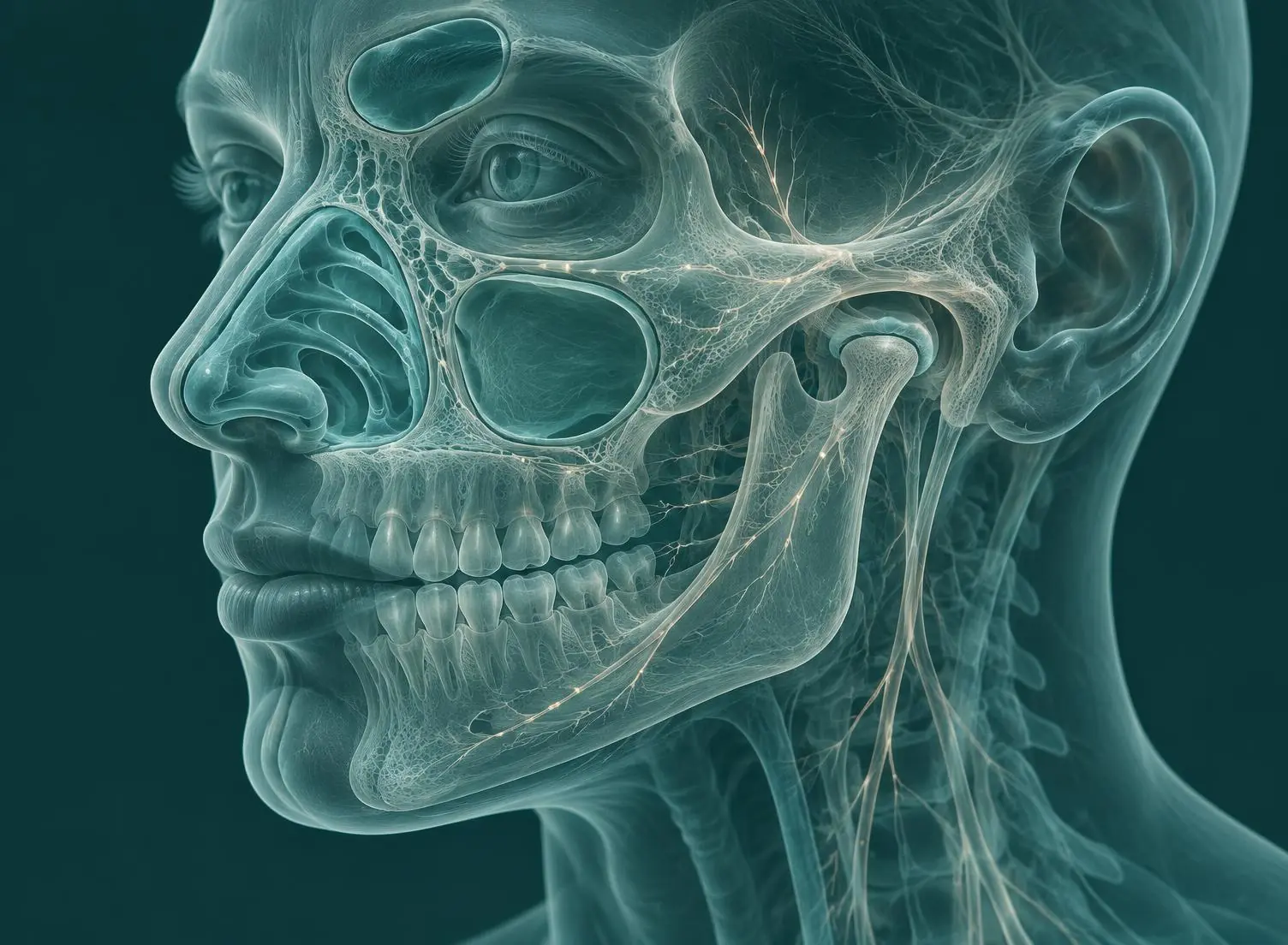 Plate 01 / Cranial & jaw anatomy
01 / Comparing models
Dental Splints & Injections
vs Nasal RX Structural Protocol
A side-by-side comparison of the conventional dental / pharmacological model and the Nasal RX structural protocol.
Primary Target
- Conventional Model (Splints / Botox)
- Teeth contact surface or muscular paralysis.
- Nasal RX Structural Protocol
- Temporal jaw socket and cranial dural alignment.
Root Cause Address
- Conventional Model (Splints / Botox)
- Buffers enamel wear; temporarily paralyses the symptom-producing muscles.
- Nasal RX Structural Protocol
- Releases sutural restriction holding the jaw socket out of alignment.
Anatomical Scope
- Conventional Model (Splints / Botox)
- Dental arch and surface muscles.
- Nasal RX Structural Protocol
- Temporal, sphenoid and dural walls.
Trigeminal Impact
- Conventional Model (Splints / Botox)
- Does not reduce mechanical nerve compression.
- Nasal RX Structural Protocol
- Decompresses CN V3 nerve branches to calm motor spasm.
Clenching Reflex
- Conventional Model (Splints / Botox)
- A plastic barrier cushions the bite.
- Nasal RX Structural Protocol
- Calms the trigeminal motor drive (CN V3).
Airway Integration
- Conventional Model (Splints / Botox)
- Rarely addresses the nasal vault.
- Nasal RX Structural Protocol
- Expands the upper nasal airflow valve.
Recovery / Safety
- Conventional Model (Splints / Botox)
- Dental downtime or injection side effects.
- Nasal RX Structural Protocol
- Zero downtime, non-surgical micro-mobilisation.
| Clinical Feature | Conventional Model (Splints / Botox) | Nasal RX Structural Protocol |
|---|---|---|
| Primary Target | Teeth contact surface or muscular paralysis. | Temporal jaw socket and cranial dural alignment. |
| Root Cause Address | Buffers enamel wear; temporarily paralyses the symptom-producing muscles. | Releases sutural restriction holding the jaw socket out of alignment. |
| Anatomical Scope | Dental arch and surface muscles. | Temporal, sphenoid and dural walls. |
| Trigeminal Impact | Does not reduce mechanical nerve compression. | Decompresses CN V3 nerve branches to calm motor spasm. |
| Clenching Reflex | A plastic barrier cushions the bite. | Calms the trigeminal motor drive (CN V3). |
| Airway Integration | Rarely addresses the nasal vault. | Expands the upper nasal airflow valve. |
| Recovery / Safety | Dental downtime or injection side effects. | Zero downtime, non-surgical micro-mobilisation. |
Clinical deep-dive library
The Full Deep-Dive Library
All long-form clinical content is preserved below. Tap any heading to expand the full spheno-temporal mechanics, articular references, FAQs and academic citations.
Why a Night Guard Alone Cannot Stop the Clicking
The temporomandibular joint is mechanically tethered to the sphenoid bone through the sphenomandibular ligament. When mechanical restrictions lock the central cranial base, the attached temporal bone rotates, shifting the glenoid fossa and altering the path of the mandibular condyle during opening.
- Sphenoid to temporal link
- The jaw socket (glenoid fossa) sits directly on the temporal bone. When the central sphenoid bones behind the nose carry mechanical tension, the attached temporal bone twists, holding the jaw socket in a sub-millimetre shift.
- Forced muscle spasm
- That sub-millimetre socket shift forces the jaw muscles into perpetual spasm, producing clicking, limited opening, and deep facial pressure.
- Trigeminal motor drive
- Dural membranes passing through the foramen ovale irritate the mandibular division of the trigeminal nerve (CN V3), driving involuntary night clenching.
Asymmetric dural tension across the sphenobasilar junction rotates the temporal bone, shifting the glenoid fossa anteriorly or laterally. This structural shift alters the trajectory of the mandibular condyle during opening, causing the fibrocartilaginous disc to displace anteriorly.
The motor nucleus of the trigeminal nerve innervates the primary muscles of mastication. Mechanical stretch applied to dural membranes passing through the foramen ovale irritates the mandibular division (CN V3), driving involuntary clenching during sleep. Releasing central dural tension quiets this hyperactive motor neuron firing.
A splint cushions the teeth, but it cannot realign a rotated temporal bone or release the dural tension driving the socket shift.
The temporal bone forms the mandibular fossa and articular eminence. Because the temporal bone articulates with the sphenoid, occiput, and parietal bones, rotational torque at the sphenobasilar junction alters the position of the glenoid fossa relative to the condyle.
This structural mismatch compresses the highly vascularised and innervated retrodiscal tissue, forcing the fibrocartilaginous disc to displace anteriorly during jaw depression. Endonasal micro-mobilisation acts upon the palatine, sphenoid, and vomer axis to restore cranial equilibrium, relieving retrodiscal compression and restoring smooth joint tracking.
Nasal RX supports the structural source of TMJ strain by releasing the cranial base tension that twists the temporal bone:
- 01
Realigning the Temporal Socket
By gently mobilising the central sphenoid anchor from within the nasal passageway, the attached temporal bone is allowed to return to a neutral orientation, decompressing the jaw socket.
- 02
Calming the Trigeminal Motor Drive
Releasing central dural tension quiets hyperactive motor neuron firing along CN V3, reducing involuntary clenching without relying on a plastic barrier.
- 03
Restoring Nasal Airway Integration
Opening the upper nasal airflow valve addresses the airway-clenching reflex that frequently accompanies chronic TMJ strain.
- 1Okeson JP. Management of Temporomandibular Disorders and Occlusion (8th ed.). St. Louis, MO: Elsevier Health Sciences; 2019.
- 2Scarr G. Biotensegrity of the temporomandibular joint: molecular and structural mechanics. J Bodyw Mov Ther. 2020;24(1):89-97.
- 3Willard FH, Vleeming A, Schuenke MD, Danneels L, Schleip R. The thoracolumbar fascia: anatomy, function and clinical considerations. J Anat. 2012;221(6):507-536.
- 4Sessle BJ. Peripheral and central mechanisms of orofacial pain and their clinical correlates. Minerva Anestesiol. 2011;77(1):179-206. (Int Rev Neurobiol. 2011;97:179-206.)
- 5Stiesch-Scholz M, Fink M, Tschernitschek H. Radiographic and MRI evaluation of spheno-temporal alignment in patients with internal derangement of the TMJ. J Oral Rehabil. 2003;30(7):712-719.
Academic references are provided for educational and context purposes. Individual clinical outcomes vary, and suitability for treatment is determined during a personal clinical consultation.
The scientific citations referenced on this website represent foundational anatomical, biomechanical, and physiological research. They are provided for educational and context purposes. Individual clinical outcomes vary, and suitability for treatment is determined during a personal clinical consultation.
Rebalance Your Jaw Mechanics Today
Schedule a comprehensive structural assessment to evaluate your cranial base mechanics, temporal alignment, and trigeminal pathways.
Request Jaw Assessment