
An illustrated research guide
Dura Mater and Referred Head Pain
Where the dura attaches, where pain has been reported during stimulation, and what the research can tell us.
Clinical lead at Cranial Solutions: Dr John Gibson, chiropractor
BMedSci (Hons Pharma), MChiro
What is the dura mater?
The dura mater is the tough outer meningeal covering of the brain and spinal cord. Inside the skull, it lines bone and forms internal folds, including the falx cerebri and tentorium cerebelli. [11]
The skull-lining periosteal layer and the inward-folding meningeal layer have different relationships to bone. A fold has free surfaces as well as attached margins; it is not glued to a single skull bone throughout its extent.
Can stimulation of the dura refer pain elsewhere in the head?
Yes. Human surgical observations have recorded pain away from the stimulated dural site. Several structures can produce overlapping pain locations, so a pain location alone cannot identify its source. [3] [5] [8]
The map reads from stimulated structure to reported pain. It cannot safely be reversed into “pain here means this dural attachment is the cause”.
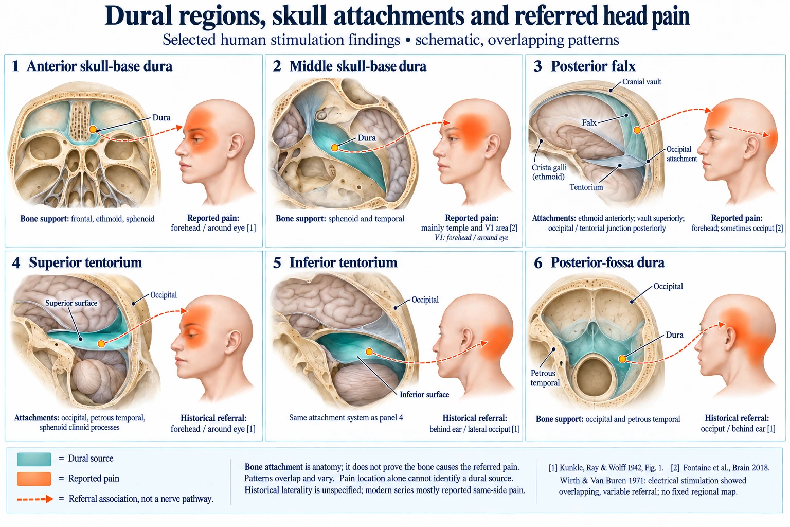
Teal marks the dural region; orange marks a reported pain area. Dashed arrows show referral associations, not traced nerve pathways. Selected patterns are schematic and overlap. [3] [8] How to cite or share this illustration.
Which skull bones are associated with the six dural regions?
The skull-base regions line bone. The falx and tentorium are internal folds supported along their attached margins. Bone attachment and pain referral are separate anatomical and observational facts. [11] [12] [13]
Bone support and attachment context Dural region Associated bones Attachment context 1. Anterior skull-base dura Frontal, ethmoid and sphenoid Lines the orbital roofs, cribriform plate and anterior skull-base portions of the sphenoid. 2. Middle skull-base dura Sphenoid and temporal bones Lines the greater sphenoid wings and temporal portions of the middle-fossa floor. 3. Posterior falx Occipital bone and the tentorial junction The whole falx also attaches to the ethmoid crista galli, frontal crest and inner midline vault. Those anterior attachments are not attachments of the posterior segment alone. 4. Superior tentorium Occipital, petrous temporal and sphenoid Anchored along occipital transverse-sinus grooves, petrous ridges and sphenoid clinoid processes. 5. Inferior tentorium The same bones as the superior surface Upper and lower surfaces belong to one fold, with the same peripheral attachment system. 6. Posterior-fossa dura Mainly occipital and temporal bones Lines the posterior cranial bowl; the anterior clival region also involves sphenoid–occipital bone.
Where has pain been reported after dural stimulation?
This table keeps the historical chart, modern mechanical-contact observations and electrical stimulation separate. Entries marked as gaps have no independently verified regional mapping in this synthesis; that does not mean the tissue is insensitive.
Historical chart descriptions summarize Figure 1 of Kunkle, Ray and Wolff (1942). Laterality is not specified for every entry. Modern mechanical-contact observations were ipsilateral in the 2018 series; electrical responses in 1971 were more variable. [3] [8] [5]
25 of 25 entries
Stimulated structure → reported pain location Structure Reported pain / gap Source & evidence Limits Anterior-fossa dural floor Fronto-orbital [3] Historical chart: directly inspectedFig.1, p.403 (PDF p.4) Historical shading is regional, not a precise boundary. Cavernous sinus Fronto-orbital [3] Historical chart: directly inspectedFig.1, p.403 (PDF p.4) Historical shading is regional, not a precise boundary. Anterior sagittal sinus / tributaries Temporal and vertex [3] Historical chart: directly inspectedFig.1, p.403 (PDF p.4) Historical shading is regional, not a precise boundary. Sagittal sinus / tributaries (segment unspecified) Fronto-orbital [3] Historical chart: directly inspectedFig.1, p.403 (PDF p.4) Historical shading is regional, not a precise boundary. Superior tentorium Fronto-orbital [3] Historical chart: directly inspectedFig.1, p.403 (PDF p.4) Historical shading is regional, not a precise boundary. Inferior tentorium Postauricular / lateral occipital [3] Historical chart: directly inspectedFig.1, p.403 (PDF p.4) Historical shading is regional, not a precise boundary. Superior transverse sinus Fronto-orbital [3] Historical chart: directly inspectedFig.1, p.403 (PDF p.4) Historical shading is regional, not a precise boundary. Inferior transverse sinus Postauricular / lateral occipital [3] Historical chart: directly inspectedFig.1, p.403 (PDF p.4) Historical shading is regional, not a precise boundary. Straight sinus Fronto-orbital and postauricular [3] Historical chart: directly inspectedFig.1, p.403 (PDF p.4) Historical shading is regional, not a precise boundary. Torcular Herophili Fronto-orbital [3] Historical chart: directly inspectedFig.1, p.403 (PDF p.4) Historical shading is regional, not a precise boundary. Sigmoid sinus Postauricular / lateral occipital [3] Historical chart: directly inspectedFig.1, p.403 (PDF p.4) Historical shading is regional, not a precise boundary. Occipital sinus Occipital / suboccipital [3] Historical chart: directly inspectedFig.1, p.403 (PDF p.4) Historical shading is regional, not a precise boundary. Posterior-fossa dural floor Postauricular and occipital [3] Historical chart: directly inspectedFig.1, p.403 (PDF p.4) Historical shading is regional, not a precise boundary. Dura at internal auditory meatus Postauricular [3] Historical chart: directly inspectedFig.1, p.403 (PDF p.4) Historical shading is regional, not a precise boundary. Middle meningeal arterial region Temporal [3] Historical chart: directly inspectedFig.1, p.403 Historical shading is regional, not a precise boundary. Skull-base dura Mainly forehead/periorbital (V1) and temple [8] Modern human mechanical contactResults p.1044; Fig.2 p.1042 See study context below. Falx (pooled) Mainly forehead/periorbital and temple [8] Modern human mechanical contactResults p.1044; Fig.2 See study context below. Posterior falx Forehead; occasional occipital referral [8] Modern human mechanical contactDiscussion p.1046 See study context below. Posterior middle-fossa dura Occasional occipital referral [8] Modern human mechanical contactDiscussion p.1046 See study context below. Frontal convexity Individual numbered map requires further visual extraction [8] Reported observation; referral not transcribedFig.2 caption, event 14; discussion p.1045 Caption notes convexity despite pooled skull-base/falx summary; do not add a 31st event. Multiple dural electrode sites V1–V3 and C1–C3; variable side [5] Human electrical stimulationIndexed original abstract; full-text maps pending No reproducible region-to-region map from skull landmarks; not a mechanical stretch test. Posterior falx; tentorium; middle-fossa floor Pain on manipulation; no new uniform referral map assigned [6] Review plus surgical casesAbstract Results; surgical case review Supports sensitivity; distinguish historical maps discussed by the review from new case observations. Anterior falx, isolated from posterior falx No separately verified referral row in this extraction [8] GapFig.2 / text: pooled falx Do not substitute an anatomical nerve territory for an observed referral. Posterior sagittal sinus, isolated Not separately resolved here [3] GapFig.1: generic/anterior labels only Generic sinus labels cannot be extended to an unlabelled posterior segment. Parietal / occipital convexity dura No adequately verified regional map here [8] GapDiscussion p.1045: sampling limitation Unmapped does not mean insensitive. On smaller screens, scroll within the table to read all columns. V1, V2 and V3 denote ophthalmic, maxillary and mandibular trigeminal territories; C1–C3 denote upper cervical nerve territories.
What do the historical and modern human studies show?
Wirth and Van Buren, 1971
Electrical stimulation through implanted electrodes in 25 patients elicited referral across trigeminal and upper cervical territories, including bilateral and contralateral responses. A consistent map could not be established from electrode positions defined by skull landmarks. The indexed abstract supports this caution; the full electrode diagrams have not been checked for this guide. [5]
Modern awake-craniotomy observations
Fontaine and colleagues collected 93 painful events in 53 patients. Thirty were reported as dural events: 23 skull-base and seven falx events. Dural stimulation was slight touch or contact during necessary surgery, not a calibrated stretching experiment. Referral was chiefly forehead/periorbital and temporal, with variation. [8]
The convexity dura had been locally anaesthetized, and sampling depended on the surgery. Event counts therefore cannot rank regional nerve density. The paper also reports non-dural pain-sensitive structures; those events must not be counted as dural events.
A 2012 case-based review and later surgical surveys and guidance provide additional clinical context. They do not create a new, uniform dural referral atlas. [6] [7] [9] [10]
What does a posterior falx or tentorial attachment mean?
The posterior falx joins the tentorium near its posterior attachment. The whole falx extends forward to the ethmoid crista galli and frontal crest and follows the inner midline vault. Upper and lower tentorial surfaces share the same attachment system. [12] [13]
An attached bone, a stimulated dural site and a perceived skin-pain area are three different things. Their anatomical relationship does not establish that bone displacement is causing an individual’s headache.
Does this explain every migraine or prove a treatment mechanism?
No. Surgical pain provocation does not establish the cause of an individual’s chronic headache or show that a treatment changes dural tension. It also does not measure what percentage of cranial pain receptors are in the dura.
Mechanical sensitivity, chemical signalling such as CGRP release, chronic headache causation and treatment outcomes need separate evidence. This guide focuses on cranial referral findings, rather than spinal tension tests or exercise prescriptions.
How can I cite or share the illustration?
Link to this research guide so readers can see the source studies, limitations and current version together. Please retain the Cranial Solutions source credit when sharing the illustration and contact us about reproduction or high-resolution teaching use.
Cranial Solutions. Dura Mater and Referred Head Pain: An Illustrated Research Guide. Illustration and literature synthesis.
https://www.cranialsolutions.nz/articles/dura-mater-referred-painThis illustration is an AI-assisted educational synthesis, not a figure from the original studies. No independent anatomical or specialist review is claimed.
Ask about illustration use →
Where can I read more about headache and neck symptoms?
Which studies and anatomical sources support this guide?
Numbered references link to the original or indexed source. The access notes identify which regional findings were checked directly and where original full-text access remains incomplete.
- Experimental studies on headache: pain-sensitive structures of the head and their significance in headache
Bronson S. Ray; Harold G. Wolff. 1940. Archives of Surgery 41(4):813–856.
Bibliographic record and later accounts checked; original full text not obtained. Regional rows below use K42 explicitly, not an asserted direct extraction of R40. - Dural headache and innervation of the dura mater
Wilder Penfield; Francis McNaughton. 1940. Archives of Neurology and Psychiatry 44:43–75.
Original full text not obtained; citation cross-checked in F18. Do not confuse with later abstracts/reprints. - Studies on headache: the mechanisms and significance of the headache associated with brain tumor
E. Charles Kunkle; Bronson S. Ray; Harold G. Wolff. 1942. Bulletin of the New York Academy of Medicine 18(6):400–422.
Original PDF inspected, including visual reading of Figure 1, printed p.403 / PDF page 4. - The tentorial nerves and localization of intracranial pain in man
William Feindel; Wilder Penfield; Francis McNaughton. 1960. Neurology 10:555–563.
Citation verified; full original mapping figures not obtained. - Referral of pain from dural stimulation in man
F. P. Wirth Jr; J. M. Van Buren. 1971. Journal of Neurosurgery 34(5):630–642.
PubMed metadata and indexed original abstract checked through Consensus. PubMed has no abstract. Full text/individual electrode diagrams not obtained. - The innervation of the cranial dura mater: neurosurgical case correlates and a review of the literature
William J. Kemp III; R. Shane Tubbs; Aaron A. Cohen-Gadol. 2012. World Neurosurgery 78(5):505–510; online 2011.
Primary abstract checked; regional historical accounts distinguished from original mapping. - Pain during awake craniotomy for brain tumor resection. Incidence, causes, consequences and management
Denys Fontaine; Fabien Almairac. 2017. Neurochirurgie 63(3):204–207.
Indexed abstract and bibliographic record checked. - Dural and pial pain-sensitive structures in humans: new inputs from awake craniotomies
Denys Fontaine; Fabien Almairac; Serena Santucci; Charlotte Fernandez; Radhouane Dallel; Johan Pallud; Michel Lanteri-Minet. 2018. Brain 141(4):1040–1048.
Full text checked, especially Methods p.1042, Results p.1044, discussion pp.1045–1046 and Figure 2 caption. - Anesthesia management for low-grade glioma awake surgery: a European Low-Grade Glioma Network survey
Arzoine et al.. 2020. Acta Neurochirurgica 162:1701–1707.
Indexed citation/abstract checked; not used for regional mapping. - Practical guidance for monitored anesthesia care during awake craniotomy
Kyeong Tae Min. 2025. Anesthesia and Pain Medicine (Seoul) 20(1):23–33.
Full-text review checked; not used as original mapping data. - Anatomy, Head and Neck, Dura Mater
StatPearls. online edition; accessed 1 November 2026. NCBI Bookshelf.
Anatomical background; not a referral-mapping experiment. - Neuroanatomy, Falx Cerebri
StatPearls. online edition; accessed 1 November 2026. NCBI Bookshelf.
Anatomical attachment context; not evidence of a treatment effect. - The Tentorium Cerebelli: A Comprehensive Review Including Its Anatomy, Embryology, and Surgical Techniques
Rabjot Rai; Joe Iwanaga; Gaffar Shokouhi; Rod J. Oskouian; R. Shane Tubbs. 2018. Cureus.
Anatomical review; source for the dural-fold attachment system.
Scope and update record
Literature and source checking: 1 November 2026. Targeted synthesis of human cranial stimulation evidence and anatomy, not an exhaustive systematic review. The table preserves overlapping findings and access gaps. This date records research preparation, not independent clinical review.
- Experimental studies on headache: pain-sensitive structures of the head and their significance in headache